A Deeper Look Into The Structure And Function Of The Knee
The knee is one of the most complex and vital joints in the human body, enabling movement while bearing the body’s weight. Understanding the anatomy of the knee can help us appreciate its function, the common injuries it can sustain, and how to maintain its health. In this blog, we delve deep into the structure and functionality of the knee joint.
Overview of the Knee Joint
The knee is a hinge joint, primarily allowing for bending and straightening motions, along with a slight degree of rotation and sliding. It connects the thigh bone (femur) to the shin bone (tibia), with the kneecap (patella) situated at the front to provide protection and leverage. The fibula, a smaller bone next to the tibia, also forms part of the knee joint but is mainly involved in muscle attachment rather than joint movement.
Key Structures of the Knee
Bones
- Femur: The upper leg bone has two rounded knobs at its lower end, known as the femoral condyles, which articulate with the tibia and patella.
- Tibia: The larger bone of the lower leg, the tibia supports most of the body’s weight. Its upper end forms a joint with the femur and patella.
- Patella: Also known as the kneecap, it is embedded within the quadriceps tendon and improves the knee’s mechanical advantage.
Ligaments
Ligaments are tough bands of tissue that connect bones to each other, providing stability to the joint.
- Anterior Cruciate Ligament (ACL): Prevents the femur from sliding backward on the tibia.
- Posterior Cruciate Ligament (PCL): Prevents the femur from sliding forward on the tibia.
- Medial Collateral Ligament (MCL): Provides stability to the inner knee.
- Lateral Collateral Ligament (LCL): Stabilizes the outer knee.
Cartilage
Cartilage cushions the joint and allows for smooth movement.
- Menisci: Two C-shaped pieces of cartilage between the femur and tibia, acting as shock absorbers and stabilizers.
- Articular Cartilage: Covers the surfaces of the bones inside the joint, reducing friction and cushioning during movement.
Muscles
Muscles are essential for the movement of the knee joint.
- Quadriceps: Located at the front of the thigh, these muscles extend the knee.
- Hamstrings: Located at the back of the thigh, they are responsible for bending the knee.
Tendons
Tendons connect muscles to bones, allowing for movement.
- Patellar Tendon: Connects the patella to the tibia and plays a crucial role in the knee’s ability to extend.
Function of the Knee
The knee’s primary function is to allow leg movement necessary for activities such as walking, running, and jumping. It supports the body’s weight, absorbs shock, and maintains balance. The unique structure of the knee, with its combination of bone, ligaments, cartilage, muscles, and tendons, is optimized for both mobility and stability.
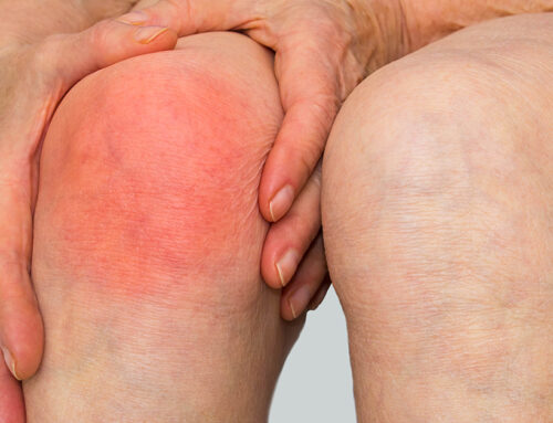
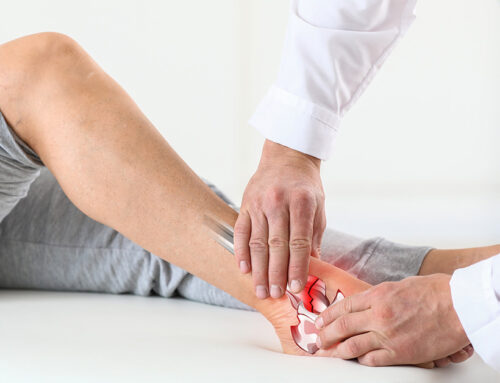
Common Knee Injuries
Due to its complexity and the loads it bears, the knee is susceptible to various injuries:
- Ligament Sprains or Tears: ACL and MCL injuries are common in athletes.
- Meniscus Tears: Often occur during sports when the knee is twisted while bearing weight.
- Patellar Tendonitis: Overuse injury often seen in runners and jumpers.
- Fractures: The patella or other bones of the knee can break during severe impacts.
Maintaining Healthy Knees
Maintaining knee health is crucial for mobility and quality of life. Regular exercise, maintaining a healthy weight, avoiding excessive stress, and proper nutrition can help sustain knee health. Strengthening the muscles around the knee and maintaining flexibility also play essential roles in supporting and stabilizing the joint.
The knee’s anatomy is designed to perform its function efficiently, balancing both mobility and stability. Understanding the knee’s complex structure helps in recognizing the importance of each component, leading to better prevention and management of knee problems. Whether you’re an athlete, fitness enthusiast, or just looking to keep your knees healthy as you age, knowing more about this vital joint can empower you to take better care of it.